Neuroscience
See neural tissues clearly
The high heterogeneity and autofluorescence of brain and neural tissue present technical challenges when using conventional imaging techniques to decipher different cell types and intricate spatial relationships in the study of neurodegenerative disease or neuro-oncology.
Imaging Mass Cytometry™ (IMC™) produces clear, easy-to-interpret signals from neural tissue, eliminating autofluorescence interference with metal-labeled antibodies.
Download neuroscience bibliography
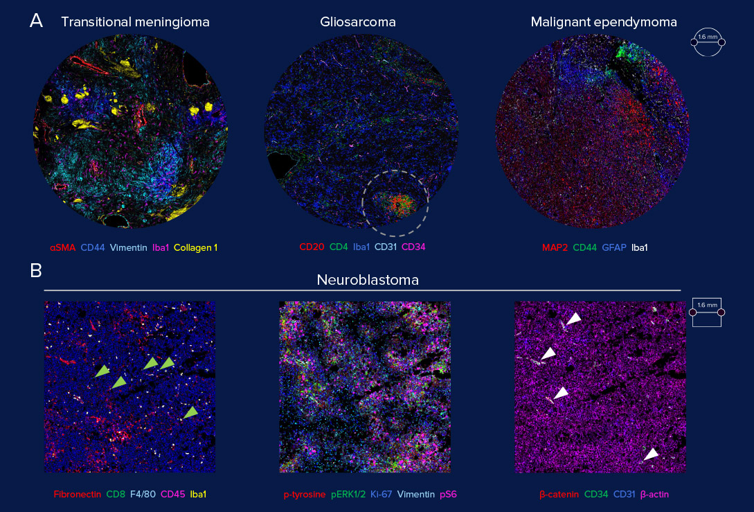
Application of neuro-oncology IMC panel on human and mouse tumors of mixed or non-glial origin
Mapping the neuro-oncological cellular landscape
Spatial phenotyping with IMC enables comprehensive assessment of the structural and cellular organization of the tumor microenvironment in neural tissues, including activation of immuno-oncological processes.
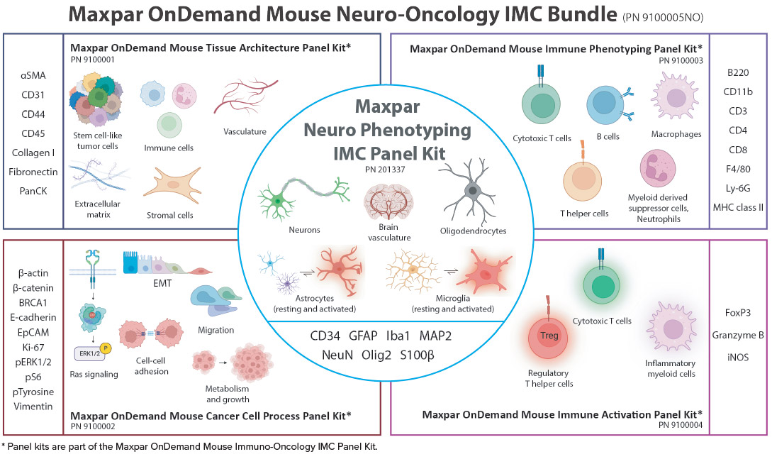
Neuro-oncology panel kits for Imaging Mass Cytometry
Identify neural cell lineages and immune cell infiltration in spatial context to better understand disease progression.
Try one of our ready-made panels or bundles or explore our comprehensive catalog of immune markers. Our easy panel design process makes integrating new markers simple.
Learn about the power of the high-plex Maxpar OnDemand Mouse Neuro-Oncology IMC Bundle to enable single-cell analysis for fast and accurate brain tumor and immune cell phenotyping.
Study neurodegeneration spatial complexity in context
Pathological proteins have emerged as a common mechanism for the progression of various neurodegenerative diseases. Discover the spatial distribution of these key proteins in neural tissues using pre-designed IMC panels for the study of neurodegenerative diseases in formalin-fixed, paraffin-embedded (FFPE) tissue sections.
Alzheimer’s disease
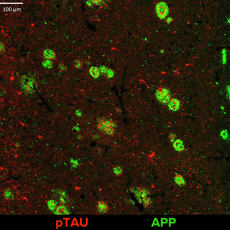 Study Alzheimer’s disease and tauopathies or amyloidopathies, such as progressive supranuclear palsy with markers targeting Tau, phosphorylated Tau (pTau) and amyloid precursor protein (APP).
Parkinson’s disease
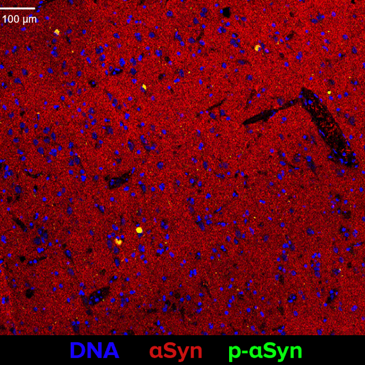 Study Parkinson’s disease or similar conditions, such as dementia with Lewy bodies with markers targeting αSynuclein (αSyn), phosphorylated αSynuclein (p-αSyn) and tyrosine hydroxylase (TH).
Multiple sclerosis
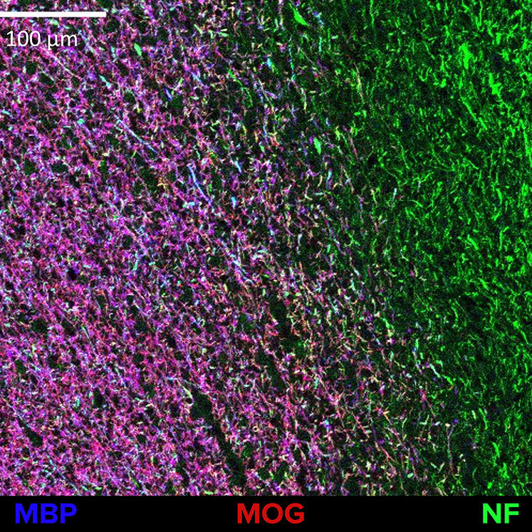 Study multiple sclerosis and other demyelinating disorders of the central nervous system with markers targeting human- and mouse-reactive myelin basic protein (MBP), myelin oligodendrocyte glycoprotein (MOG) and pan-axonal neurofilament (NF) protein.
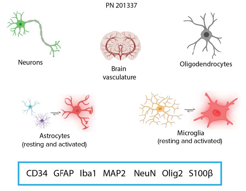
Add neurophenotyping markers for deeper insights
Capture a more complete view of neurodegeneration by combining any neurodegeneration panel with the Maxpar Neuro Phenotyping IMC Panel Kit. Study immune cell infiltration and activation states of complex immunological markers.
Add single-cell analysis to your panel with improved nucleus and plasma membrane demarcation.


Webinar: Spatial Dissection Of Tumor Immune Microenvironment
Daniela Quail, PhD
Assistant Professor, Rosalind and Morris Goodman Cancer Institute and Department of Physiology, McGill University
Quail describes the use of Imaging Mass Cytometry to understand immune dynamics in tumors – first in the brain tumor microenvironment to identify new cell populations that might help understand tumor biology and second in non-small-cell lung cancer patients to predict recurrence and survival outcomes.
Unless explicitly and expressly stated otherwise, all products are provided for Research Use Only, not for use in diagnostic procedures. Find more information here.